| DocDalgatov | Дата: Понедельник, 22 Апреля 13, 17:11:09 | Сообщение # 1 |
|
Администратор
Группа: Администратор
Сообщений: 437
Репутация: 4
Статус:  | http://www.nature.com/mp/journal/v10/n10/full/4001711a.html
Выдержки из статьи
Molecular Psychiatry (2005) 10, 900–919. doi:10.1038/sj.mp.4001711; published online 12 July 2005
Mitochondrial dysfunction in bipolar disorder: evidence from magnetic resonance spectroscopy research
C Stork and P F Renshaw
Brain Imaging Center, McLean Hospital, Belmont, MA, USA
Correspondence: C Stork, Brain Imaging Center, McLean Hospital, 115 Mill St, Belmont, MA 02478, USA. E-mail: cstork@mclean.harvard.edu
Received 2 May 2005; Revised 3 June 2005; Accepted 8 June 2005; Published online 12 July 2005.
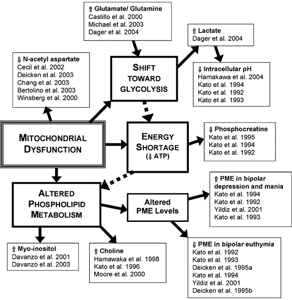
"Magnetic resonance spectroscopy (MRS) affords a noninvasive window on in vivo brain chemistry and, as such, provides a unique opportunity to gain insight into the biochemical pathology of bipolar disorder. Studies utilizing proton (1H) MRS have identified changes in cerebral concentrations of N-acetyl aspartate, glutamate/glutamine, choline-containing compounds, myo-inositol, and lactate in bipolar subjects compared to normal controls, while studies using phosphorus (31P) MRS have examined additional alterations in levels of phosphocreatine, phosphomonoesters, and intracellular pH. We hypothesize that the majority of MRS findings in bipolar subjects can be fit into a more cohesive bioenergetic and neurochemical model of bipolar illness that is both novel and yet in concordance with findings from complementary methodological approaches. In this review, we propose a hypothesis of mitochondrial dysfunction in bipolar disorder that involves impaired oxidative phosphorylation, a resultant shift toward glycolytic energy production, a decrease in total energy production and/or substrate availability, and altered phospholipid metabolism.
Reduced N-acetyl-aspartate in bipolar disorder: marker of mitochondrial dysfunction.
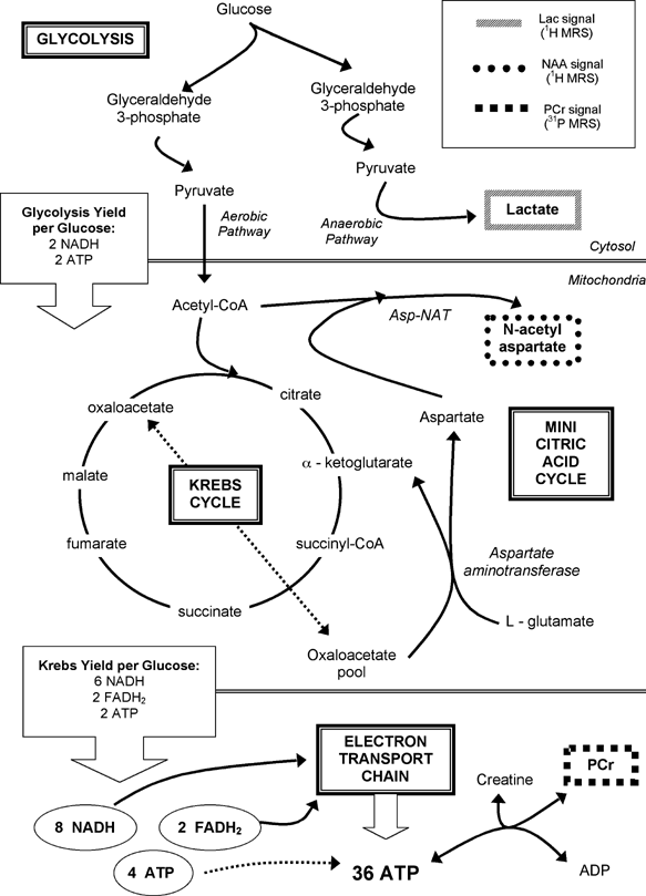
After early research with rat brain mitochondria suggested that NAA is of mitochondrial origin,6 Truckenmiller et al7 demonstrated that NAA is synthesized in mitochondria by the membrane-bound enzyme L-aspartate N-acetyltransferase, a catalyst that is found only in the brain.
Consequently, although reductions in NAA concentration were formerly thought to indicate neuronal death,12 recent research has suggested that decreased levels of NAA are more accurately consistent with impaired mitochondrial energy production.
Researchers have also investigated whether observations of reduced NAA in bipolar subjects may be due to various treatments for the disorder, in particular lithium administration.
However, several trials have suggested that lithium administration actually increases NAA levels in the brain.
Even in contrast with these results, studies led by Brambilla et al26 in healthy individuals and by Friedman et al27 in bipolar subjects have found that lithium administration does not appear to be associated with significant differences in NAA concentration. Thus, there remains a lack of MRS evidence that lithium administration is associated with the reduced NAA levels repeatedly observed in bipolar subjects.
Specifically, findings of both decreased pHi and increased levels of lactate in bipolar subjects suggest a shift away from oxidative phosphorylation toward glycolysis, thus reducing efficiency and reducing total energy output.
As reported by Kato and co-workers, at the present time, reduced pHi has only been observed in pathological states that are known to arise from ischemic insult to the brain, including white matter hyperintensities,35 acute stroke,36 and subarachnoid hemorrhage.
Consequently, findings of decreased pHi in bipolar subjects compared to normal controls suggest that impaired mitochondrial function may be an integral component of bipolar illness.
Instead, Kato and Kato52 have hypothesized that the decreased pHi observed in euthymic bipolar subjects is a trait of the illness itself. Specifically, these researchers have suggested that manic and depressive states may be at least partially caused by the brain's attempts to correct this pH imbalance by causing an overactivation of monoaminergic systems, which has been known to increase pHi at least in hippocampal neurons.
Reductions in cerebral pHi such as those observed in bipolar subjects have also frequently been linked to increased levels of lactate, a condition that is known to result from mitochondrial dysfunction.
When mitochondrial function is inhibited, rendering the respiratory chain of cellular metabolism unavailable, the only mode of energy production available to the cell is anaerobic glycolysis.
An increase in lactate concentration therefore suggests an increase in the rate of glycolysis, and consequently also implies an inhibition of the mitochondrial oxidative phosphorylation process; lactate is known to accumulate only when oxidative phosphorylation is unable to meet energy requirements and the cell is forced to rely on the glycolytic process.
If cells are unable to meet such increased energy requirements through mitochondria-based oxidative phosphorylation, rates of glycolysis would increase, thus causing the increased levels of lactate and decreased pHi observed in studies of bipolar disorder.
Interestingly, parallel findings of increased glutamate, glutamine, and lactate have lent support to theories of mitochondrial dysfunction in Huntington's disease (HD)
Reduced PCr concentrations have also been reported in migraine, which has been postulated to be associated with abnormal mitochondrial function.
A similar study by Kato et al,49only 1 year earlier, also reported significantly decreased whole brain PCr in bipolar II patients, but, interestingly, not bipolar I patients, relative to normal controls.
Nonetheless, these alterations of PCr in bipolar disorder are consistent with some degree of mitochondrial involvement in both the illness and its treatment.
Further support for a theory of mitochondrial dysfunction in bipolar disorder has arisen from MRS studies of phospholipid metabolism in bipolar subjects. In a normal brain cell, the synthesis and maintenance of the cell membrane require between 10 and 15% of net brain ATP production (Figure 3).79 If less energy is produced in the cell overall, it is likely that aspects of phospholipid metabolism, including de novo phospholipid biosynthesis, would also be impaired. Several MRS studies have indicated that phospholipid metabolism is indeed abnormal in bipolar patients, as evidenced by alterations in levels of Cho, mI, inositol monophosphates, and PMEs (Tables 6, 7, 8, 9 and 10). Accordingly, we hypothesize that these alterations are the direct result of energy shortages caused by mitochondrial dysfunction.
However, additional MRS investigations of both the hippocampus,5, 21 and DLPFC2 have reported finding no significant differences in Cho/Cr + PCr or absolute Cho levels between bipolar subjects and controls. Studies of children with bipolar disorder have similarly noted no significant differences in Cho concentrations between bipolar and control subjects.20, 30 Nonetheless, it remains possible that findings of increased Cho concentration in bipolar subjects may be indicative of impaired phospholipid metabolism and thus also of mitochondrial dysfunction.several MRS studies have reported little to no association between elevated brain Cho concentration and lithium treatment (Table 7). A study by Sharma et al23 reported finding higher Cho/Cr + PCr in the basal ganglia region of bipolar subjects taking lithium compared to normal controls, but a more recent study by Wu et al90 contradicted these results by reporting that lithium-treated bipolar patients had significantly lower Cho/Cr + PCr in the temporal lobe than healthy control subjects. Consequently, if lithium administration does exert a significant effect on brain Cho metabolism, this effect remains to be clarified.
One 1998 study by Kato et al48 did not find a difference in PME levels between euthymic bipolar subjects and controls, but had an extremely small sample group of only seven subjects.
Individual studies by Kato et al have reported significantly elevated PME levels in both depressed47 and manic46 bipolar subjects compared to those in euthymia, and the later meta-analysis by Yildiz et al113 also found significantly increased PME levels in depressed vs euthymic bipolar subjects. Like measurements of altered pHi in bipolar subjects (Table 3), these state-dependent abnormalities in PME concentration may be the result of the brain's attempts to normalize membrane metabolic processes that are impaired due to mitochondrial dysfunction.
A number of PET studies have demonstrated that cerebral glucose metabolic rates are significantly decreased in bipolar subjects in depressed or mixed mood states.
Although PET research cannot identify the etiology of these metabolic differences in bipolar patients, it is possible that they are related to the mitochondrial dysfunction suggested by MRS studies.
Investigations of mitochondria-based neurological disorders, including HD, Parkinson's disease, amyotrophic later sclerosis (ALS), and mitochondrial cytopathies, have led to the development of a number of therapies that target cellular energy dysfunction.
Studies of HD, an illness with an MRS profile similar to that of bipolar disorder (increased concentrations of lactate, glutamate, and Cho, as well as decreased NAA levels136), have found that Cr supplementation in particular can significantly increase brain concentrations of Cr and ATP to normal levels, thus exerting a neuroprotective effect and slowing the process of brain atrophy.137, 138
Additional studies of Parkinson's disease and TBI have also investigated the potential use of compounds that may improve cellular energy function, such as coenzyme Q10, Gingko biloba, nicotinamide, and lipoic acid, but evidence as to the therapeutic efficacy of these treatments remains extremely limited.135, 139, 140
Yet if bipolar disorder can be viewed in terms of its possible basis in mitochondrial dysfunction, such therapies have the potential to ameliorate aspects of the disease that are only minimally addressed by current treatment methods".
|
| |
| |
| DocDalgatov | Дата: Понедельник, 22 Апреля 13, 17:54:32 | Сообщение # 2 |
|
Администратор
Группа: Администратор
Сообщений: 437
Репутация: 4
Статус:  | 

|
| |
| |
| DocDalgatov | Дата: Понедельник, 22 Апреля 13, 17:56:14 | Сообщение # 3 |
|
Администратор
Группа: Администратор
Сообщений: 437
Репутация: 4
Статус:  | Ошибочная привязка к БАР. Словосочетание "bipolar disorder" заменяем на "панические атаки".
|
| |
| |
| Иринa | Дата: Среда, 24 Апреля 13, 12:28:28 | Сообщение # 4 |
|
Активный пользователь
Группа: Пользователи
Сообщений: 292
Репутация: 5
Статус:  | А что, при БАР, разве с митохондриями всё в порядке?
|
| |
| |
| DocDalgatov | Дата: Среда, 24 Апреля 13, 13:05:04 | Сообщение # 5 |
|
Администратор
Группа: Администратор
Сообщений: 437
Репутация: 4
Статус:  | Дело не про "всё в порядке". Дело о том, что не это патогенез БАР.
Оффтоп удалён.
|
| |
| |
| Иринa | Дата: Среда, 15 Мая 13, 15:36:46 | Сообщение # 6 |
|
Активный пользователь
Группа: Пользователи
Сообщений: 292
Репутация: 5
Статус:  | При БАР с митохондриями всё впорядке? А почему тогда при униполярной депрессии митохондрии от демотивации гибнут? Неврастения ведь просто так не бывает, она лишь начинается на фоне депрессии. А если при БАР слишком длинная депрессивная фаза, то почему бы митохондриям не истощится от демотивации? А маниакальная фаза, это разве не попытка гипоталамусу восстановить активность рабоы мозга? Разве при БАР гипоталамус ни разгоняетработу ядер шва? Неужели от этой эмоциональной лабильности митохондрии не истощатся? И депрессия и мания- это всё ненормально для работы мозга, поэтому, скорее всего, митохондрии ЦНС испытывают перегрузку при любой психической паталогии. Добавлено (15.05.2013, 15:36:46)
---------------------------------------------
При любой психической патологии бывают панические атаки. При ГТР, ОКР, но и при шизофрении бывают, и при БАР тоже, так как все психиатрическе круги вложены один в другой, а самый малый круг - истощаемость психической деятельности и эмоциональные гиперстенические расстройства. Вобщем КАЖДУЮ психическую паталогию сопровождает неврастения. Т.е. неврастения бывает изолированной или вместе с другими синдромами.
|
| |
| |